Next Issue
Volume 13, September
Previous Issue
Volume 13, July


Biosensors, Volume 13, Issue 8 (August 2023) – 77 articles

Cover Story (view full-size image): 
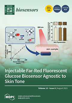
An injectable far-red fluorescence intensity-based glucose sensing assay based on solvatochromism, the reversible change in the emission spectrum dependent on the polarity of the solvent, was designed and created with a composition of PEGylated ConA and Cy5.5-labeled mannotetraose. The assay shows a linear change in fluorescence intensity at peak emission within the physiological glucose range (25 - 350 mg/dL) with a percent MARD of 12.7%. A comparison of percent change in the peak emission intensity of the assay placed beneath rat skin samples with three pigmentations (light, medium, and dark) of the same thickness (~2.1 mm), without glucose present, showed the percent change in peak emission intensity was not significantly different (p < 0.05) for the different pigmentations. Illustration created with BioRender.com. View this paper
  • Issues are regarded as officially published after their release is announced to the table of contents alert mailing list.
  • You may sign up for e-mail alerts to receive table of contents of newly released issues.
  • PDF is the official format for papers published in both, html and pdf forms. To view the papers in pdf format, click on the "PDF Full-text" link, and use the free Adobe Readerexternal link to open them.
Order results
Result details
Section
Select all
Export citation of selected articles as:
Review
Insole Systems for Disease Diagnosis and Rehabilitation: A Review
Biosensors 2023, 13(8), 833; https://doi.org/10.3390/bios13080833 - 21 Aug 2023
Viewed by 322
Abstract
Some chronic diseases, including Parkinson’s disease (PD), diabetic foot, flat foot, stroke, elderly falling, and knee osteoarthritis (KOA), are related to orthopedic organs, nerves, and muscles. The interaction of these three parts will generate a comprehensive result: gait. Furthermore, the lesions in these [...] Read more.
Some chronic diseases, including Parkinson’s disease (PD), diabetic foot, flat foot, stroke, elderly falling, and knee osteoarthritis (KOA), are related to orthopedic organs, nerves, and muscles. The interaction of these three parts will generate a comprehensive result: gait. Furthermore, the lesions in these regions can produce abnormal gait features. Therefore, monitoring the gait features can assist medical professionals in the diagnosis and analysis of these diseases. Nowadays, various insole systems based on different sensing techniques have been developed to monitor gait and aid in medical research. Hence, a detailed review of insole systems and their applications in disease management can greatly benefit researchers working in the field of medical engineering. This essay is composed of the following sections: the essay firstly provides an overview of the sensing mechanisms and parameters of typical insole systems based on different sensing techniques. Then this essay respectively discusses the three stages of gait parameters pre-processing, respectively: pressure reconstruction, feature extraction, and data normalization. Then, the relationship between gait features and pathogenic mechanisms is discussed, along with the introduction of insole systems that aid in medical research; Finally, the current challenges and future trends in the development of insole systems are discussed. Full article
(This article belongs to the Section Biosensors and Healthcare)
Show Figures

Figure 1

Article
Coupling Capillary-Driven Microfluidics with Lateral Flow Immunoassay for Signal Enhancement
Biosensors 2023, 13(8), 832; https://doi.org/10.3390/bios13080832 - 21 Aug 2023
Viewed by 363
Abstract
Microfluidics has emerged as a versatile technology that is applied to enhance the performance of analytical techniques, among others. Pursuing this, we present a capillary-driven microfluidic device that improves the sensitivity of lateral flow immunoassay rapid tests thanks to offering an automated washing [...] Read more.
Microfluidics has emerged as a versatile technology that is applied to enhance the performance of analytical techniques, among others. Pursuing this, we present a capillary-driven microfluidic device that improves the sensitivity of lateral flow immunoassay rapid tests thanks to offering an automated washing step. A novel multilevel microfluidic chip was 3D-printed with a photocurable black resin, sealed by an optically clear pressure-sensitive adhesive, and linked to the lateral flow strip. To depict the efficacy of microfluidics and the washing step, cortisol was measured quantitatively within the proposed device. Measuring cortisol levels is a way to capture physiological stress responses. Among biofluids, saliva is less infectious and easier to sample than others. However, higher sensitivity is demanded because the salivary cortisol concentrations are much lower than in blood. We carried out a competitive lateral flow immunoassay protocol with the difference that the microfluidic device applies an automated washing step after the sample is drained downstream. It washes the trapped quantum-dot-labeled antibodies out from nitrocellulose, diminishing background noise as these are bonded to cortisols and not to the immobilized receptors. Fluorescence spectroscopy, as a high-precision analysis, was successfully applied to determine clinically relevant salivary cortisol concentrations within a buffer quantitatively. The microfluidic design relied on a 3D valve that avoids reagent cross-contamination. This cross-contamination could make the washing buffer impure and undesirably dilute the sample. The proposed device is cost-effective, self-powered, robust, and ideal for non-expert users. Full article
(This article belongs to the Section Nano- and Micro-Technologies in Biosensors)
Show Figures

Figure 1

Article
A Miniature Biomedical Sensor for Rapid Detection of Schistosoma japonicum Antibodies
Biosensors 2023, 13(8), 831; https://doi.org/10.3390/bios13080831 - 20 Aug 2023
Viewed by 393
Abstract
Schistosomiasis, typically characterized by chronic infection in endemic regions, has the potential to affect liver tissue and pose a serious threat to human health. Detecting and screening for this disease early on is crucial for its prevention and control. However, existing methods encounter [...] Read more.
Schistosomiasis, typically characterized by chronic infection in endemic regions, has the potential to affect liver tissue and pose a serious threat to human health. Detecting and screening for this disease early on is crucial for its prevention and control. However, existing methods encounter challenges such as low sensitivity, time-consuming processes, and complex sample handling. To address these challenges, we report a soluble egg antigen (SEA)-based functionalized gridless and meander-type AlGaN/GaN high electron mobility transistors (HEMT) sensor for the highly sensitive detection of antibodies to Schistosoma japonicum. Immobilization of the self-assembled membrane on the gate surface was verified using a semiconductor parameter analyzer, scanning electron microscope (SEM), and atomic force microscopy (AFM). The developed biosensor demonstrates remarkable performance in detecting anti-SEA, exhibiting a linear concentration range of 10 ng/mL to 100 μg/mL and a sensitivity of 0.058 mA/log (ng/mL). It also exhibits similar excellent performance in serum systems. With advantages such as rapid detection, high sensitivity, miniaturization, and label-free operation, this biosensor can fulfill the requirements for blood defense. Full article
(This article belongs to the Section Biosensors and Healthcare)
Show Figures

Figure 1

Article
Magnetic Molecularly Imprinted Chitosan Combined with a Paper-Based Analytical Device for the Smartphone Discrimination of Tryptophan Enantiomers
Biosensors 2023, 13(8), 830; https://doi.org/10.3390/bios13080830 - 20 Aug 2023
Viewed by 317
Abstract
The separation of enantiomers plays a critical role in pharmaceutical development, ensuring therapeutic efficacy, safety, and patent protection. It enables the production of enantiopure drugs and enhances our understanding of the properties of chiral compounds. In this study, a straightforward and effective chiral [...] Read more.
The separation of enantiomers plays a critical role in pharmaceutical development, ensuring therapeutic efficacy, safety, and patent protection. It enables the production of enantiopure drugs and enhances our understanding of the properties of chiral compounds. In this study, a straightforward and effective chiral detection strategy was developed for distinguishing between tryptophan (TRP) enantiomers. The approach involved the preparation of a magnetic molecularly imprinted chitosan (MMIC) for preparation of the sample, which was combined with a nitrocellulose membrane (a paper-based analytical device, PAD) integrated with D-TRP covalently grafted with polymethacrylic acid (PAD-PMA_D-TRP). Discriminating between the TRP enantiomers was achieved using AuNPs as a colorimetric probe. Indeed, the presence of D-TRP rapidly induced the aggregation of AuNPs due to its strong affinity to PAD-PMA_D-TRP, resulting in a noticeable change in the color of the AuNPs from red to purple. On the other hand, L-TRP did not induce any color changes. The chiral analysis could be easily performed with the naked eye and/or a smartphone. The developed method exhibited a detection limit of 3.3 µM, and it was successfully applied to detect TRP in serum samples, demonstrating good recovery rates. The proposed procedure is characterized by its simplicity, cost-effectiveness, rapidity, and ease of operation. Full article
(This article belongs to the Special Issue Smartphone-Based Biosensing and Diagnostics)
Show Figures

Figure 1

Article
Voltammetric Sensing of Nifedipine Using a Glassy Carbon Electrode Modified with Carbon Nanofibers and Gold Nanoparticles
Biosensors 2023, 13(8), 829; https://doi.org/10.3390/bios13080829 - 19 Aug 2023
Viewed by 276
Abstract
Nifedipine, a widely utilized medication, plays a crucial role in managing blood pressure in humans. Due to its global prevalence and extensive usage, close monitoring is necessary to address this widespread concern effectively. Therefore, the development of an electrochemical sensor based on a [...] Read more.
Nifedipine, a widely utilized medication, plays a crucial role in managing blood pressure in humans. Due to its global prevalence and extensive usage, close monitoring is necessary to address this widespread concern effectively. Therefore, the development of an electrochemical sensor based on a glassy carbon electrode modified with carbon nanofibers and gold nanoparticles in a Nafion® film was performed, resulting in an active electrode surface for oxidation of the nifedipine molecule. This was applied, together with a voltammetric methodology, for the analysis of nifedipine in biological and environmental samples, presenting a linear concentration range from 0.020 to 2.5 × 10⁻⁶ µmol L−1 with a limit of detection 2.8 nmol L−1. In addition, it presented a good recovery analysis in the complexity of the samples, a low deviation in the presence of interfering potentials, and good repeatability between measurements. Full article
(This article belongs to the Special Issue Electrochemical Sensors and Biosensors for Biomedical Applications)
Show Figures

Graphical abstract

Article
A Facile Method for the Fabrication of the Microneedle Electrode and Its Application in the Enzymatic Determination of Glutamate
Biosensors 2023, 13(8), 828; https://doi.org/10.3390/bios13080828 - 18 Aug 2023
Viewed by 404
Abstract
Herein, a simple method has been used in the fabrication of a microneedle electrode (MNE). To do this, firstly, a commercial self-dissolving microneedle patch has been used to make a hard-polydimethylsiloxane-based micro-pore mold (MPM). Then, the pores of the MPM were filled with [...] Read more.
Herein, a simple method has been used in the fabrication of a microneedle electrode (MNE). To do this, firstly, a commercial self-dissolving microneedle patch has been used to make a hard-polydimethylsiloxane-based micro-pore mold (MPM). Then, the pores of the MPM were filled with the conductive platinum (Pt) paste and cured in an oven. Afterward, the MNE made of platinum (Pt-MNE) was characterized using cyclic voltammetry (CV), electrochemical impedance spectroscopy (EIS), and scanning electron microscopy (SEM). To prove the electrochemical applicability of the Pt-MNE, the glutamate oxidase enzyme was immobilized on the surface of the electrode, to detect glutamate, using the cyclic voltammetry (CV) and chronoamperometry (CA) methods. The obtained results demonstrated that the fabricated biosensor could detect a glutamate concentration in the range of 10–150 µM. The limits of detection (LODs) (three standard deviations of the blank/slope) were also calculated to be 0.25 µM and 0.41 µM, using CV and CA, respectively. Furthermore, the Michaelis–Menten constant (KMapp) of the biosensor was calculated to be 296.48 µM using a CA method. The proposed biosensor was finally applied, to detect the glutamate concentration in human serum samples. The presented method for the fabrication of the mold signifies a step further toward the fabrication of a microneedle electrode. Full article
(This article belongs to the Special Issue Biochips and Biosensors for Health-Care and Diagnostics)
Show Figures

Figure 1

Communication
Discrimination and Quantification of Glutathione by Cu+-Based Nanozymes
Biosensors 2023, 13(8), 827; https://doi.org/10.3390/bios13080827 - 17 Aug 2023
Viewed by 354
Abstract
Glutathione (GSH) is the most abundant low-molecular-weight biological thiol in vivo and has been linked to several diseases. The accurate quantification of GSH is therefore crucial for disease diagnosis and monitoring. In this study, we prepared self-assembled Cu(I)-Cys (cysteine) nanozymes through a two-step [...] Read more.
Glutathione (GSH) is the most abundant low-molecular-weight biological thiol in vivo and has been linked to several diseases. The accurate quantification of GSH is therefore crucial for disease diagnosis and monitoring. In this study, we prepared self-assembled Cu(I)-Cys (cysteine) nanozymes through a two-step procedure. The Cu(I)-Cys nanoparticles exhibited peroxidase-mimicking activity. Upon the addition of H2O2, they were able to oxidize 3,3,5,5-tetramethylbenzidine (TMB) into oxTMB, resulting in a measurable increase in UV-Vis absorption at 655 nm. However, in the presence of GSH, oxTMB was reduced back to TMB, leading to a decrease in UV-Vis absorption at 655 nm. By utilizing these changes in the absorption intensity, we achieved the sensitive detection of GSH with a detection limit of 2.13 μM. Moreover, taking advantage of the different peroxidase-mimicking activities of Cu(I)-Cys nanoparticles at various pH values, a sensor array with Cu(I)-Cys nanoparticles at pH 4 and pH 5 was constructed. The discrimination of GSH among Cys and ascorbic acid was achieved and the practicability of the sensor array in human serum was validated. This novel approach holds significant promise for the precise discrimination and quantification of GSH and its potential applications in disease diagnosis and therapeutics. Full article
(This article belongs to the Section Biosensors and Healthcare)
Show Figures

Figure 1

Article
Development and Evaluation of a Low-Cost Triglyceride Quantification Enzymatic Biosensor Using an Arduino-Based Microfluidic System
Biosensors 2023, 13(8), 826; https://doi.org/10.3390/bios13080826 - 17 Aug 2023
Viewed by 436
Abstract
Overweight and obesity promote diabetes and heart disease onset. Triglycerides are key biomarkers for cardiovascular disease, strokes, and other health issues. Scientists have devised methods and instruments for the detection of these molecules in liquid samples. In this study, an enzymatic biosensor was [...] Read more.
Overweight and obesity promote diabetes and heart disease onset. Triglycerides are key biomarkers for cardiovascular disease, strokes, and other health issues. Scientists have devised methods and instruments for the detection of these molecules in liquid samples. In this study, an enzymatic biosensor was developed using an Arduino-based microfluidic platform, wherein a lipolytic enzyme was immobilized on an ethylene-vinyl acetate polymer through physical adsorption. This low-cost optical biosensor employed a spectrophotometric transducer and was assessed in liquid samples to indirectly detect triglycerides and fatty acids using p-nitrophenol as an indicator. The average triglyceride level detected in the conducted experiments was 47.727 mg/dL. The biosensor exhibited a percentage of recovery of 81.12% and a variation coefficient of 0.791%. Furthermore, the biosensor demonstrated the ability to detect triglyceride levels without the need for sample dilution, ranging from 7.6741 mg/dL to 58.835 mg/dL. This study successfully developed an efficient and affordable enzymatic biosensor prototype for triglyceride and fatty acid detection. The lipolytic enzyme immobilization on the polymer substrate provided a stable and reproducible detection system, rendering this biosensor an exciting option for the detection of these molecules. Full article
(This article belongs to the Special Issue Advances in Biosensors for Health-Care and Diagnostics)
Show Figures

Figure 1

Article
Nanosphere Lithography-Enabled Hybrid Ag-Cu Surface-Enhanced Raman Spectroscopy Substrates with Enhanced Absorption of Excitation Light
Biosensors 2023, 13(8), 825; https://doi.org/10.3390/bios13080825 - 17 Aug 2023
Viewed by 285
Abstract
We demonstrated a low-cost, highly sensitive hybrid Ag-Cu substrate with enhanced absorption for the excitation laser beam via the nanosphere lithography technique. The hybrid Ag-Cu surface-enhanced Raman spectroscopy (SERS) substrate consists of a Cu nanoarray covered with Ag nanoparticles. The geometry of the [...] Read more.
We demonstrated a low-cost, highly sensitive hybrid Ag-Cu substrate with enhanced absorption for the excitation laser beam via the nanosphere lithography technique. The hybrid Ag-Cu surface-enhanced Raman spectroscopy (SERS) substrate consists of a Cu nanoarray covered with Ag nanoparticles. The geometry of the deposited Cu nanoarray is precisely determined through a self-assembly nanosphere etching process, resulting in optimized absorption for the excitation laser beam. Further Raman enhancement is achieved by incorporating plasmonic hotspots formed by dense Ag nanoparticles, grown by immersing the prepared Cu nanoarray in a silver nitrate solution. The structural design enables analytical enhancement factor of hybrid Ag-Cu SERS substrates of 1.13 × 10⁵. The Ag-Cu SERS substrates exhibit a highly sensitive and reproducible SERS activity, with a low detection limit of 10−13 M for Rhodamine 6G detection and 10−9 M for 4,4′-Bipyridine. Our strategy could pave an effective and promising approach for SERS-based rapid detection in biosensors, environmental monitoring and food safety. Full article
(This article belongs to the Special Issue Smart Materials for Chemical and Biosensing)
Show Figures

Figure 1

Article
A Critical Study on DNA Probes Attached to Microplate for CRISPR/Cas12 Trans-Cleavage Activity
Biosensors 2023, 13(8), 824; https://doi.org/10.3390/bios13080824 - 17 Aug 2023
Viewed by 363
Abstract
CRISPR/Cas12-based biosensors are emerging tools for diagnostics. However, their application of heterogeneous formats needs the efficient detection of Cas12 activity. We investigated DNA probes attached to the microplate surface and cleaved by Cas12a. Single-stranded (ss) DNA probes (19 variants) and combined probes with [...] Read more.
CRISPR/Cas12-based biosensors are emerging tools for diagnostics. However, their application of heterogeneous formats needs the efficient detection of Cas12 activity. We investigated DNA probes attached to the microplate surface and cleaved by Cas12a. Single-stranded (ss) DNA probes (19 variants) and combined probes with double-stranded (ds) and ssDNA parts (eight variants) were compared. The cleavage efficiency of dsDNA-probes demonstrated a bell-shaped dependence on their length, with a cleavage maximum of 50%. On the other hand, the cleavage efficiency of ssDNA probes increased monotonously, reaching 70%. The most effective ssDNA probes were integrated with fluorescein, antibodies, and peroxidase conjugates as reporters for fluorescent, lateral flow, and chemiluminescent detection. Long ssDNA probes (120–145 nt) proved the best for detecting Cas12a trans-activity for all of the tested variants. We proposed a test system for the detection of the nucleocapsid (N) gene of SARS-CoV-2 based on Cas12 and the ssDNA-probe attached to the microplate surface; its fluorescent limit of detection was 0.86 nM. Being united with pre-amplification using recombinase polymerase, the system reached a detection limit of 0.01 fM, thus confirming the effectiveness of the chosen ssDNA probe for Cas12-based biosensors. Full article
Show Figures

Figure 1

Review
Electrochemical Wearable Biosensors and Bioelectronic Devices Based on Hydrogels: Mechanical Properties and Electrochemical Behavior
Biosensors 2023, 13(8), 823; https://doi.org/10.3390/bios13080823 - 15 Aug 2023
Viewed by 440
Abstract
Hydrogel-based wearable electrochemical biosensors (HWEBs) are emerging biomedical devices that have recently received immense interest. The exceptional properties of HWEBs include excellent biocompatibility with hydrophilic nature, high porosity, tailorable permeability, the capability of reliable and accurate detection of disease biomarkers, suitable device–human interface, [...] Read more.
Hydrogel-based wearable electrochemical biosensors (HWEBs) are emerging biomedical devices that have recently received immense interest. The exceptional properties of HWEBs include excellent biocompatibility with hydrophilic nature, high porosity, tailorable permeability, the capability of reliable and accurate detection of disease biomarkers, suitable device–human interface, facile adjustability, and stimuli responsive to the nanofiller materials. Although the biomimetic three-dimensional hydrogels can immobilize bioreceptors, such as enzymes and aptamers, without any loss in their activities. However, most HWEBs suffer from low mechanical strength and electrical conductivity. Many studies have been performed on emerging electroactive nanofillers, including biomacromolecules, carbon-based materials, and inorganic and organic nanomaterials, to tackle these issues. Non-conductive hydrogels and even conductive hydrogels may be modified by nanofillers, as well as redox species. All these modifications have led to the design and development of efficient nanocomposites as electrochemical biosensors. In this review, both conductive-based and non-conductive-based hydrogels derived from natural and synthetic polymers are systematically reviewed. The main synthesis methods and characterization techniques are addressed. The mechanical properties and electrochemical behavior of HWEBs are discussed in detail. Finally, the prospects and potential applications of HWEBs in biosensing, healthcare monitoring, and clinical diagnostics are highlighted. Full article
Show Figures

Figure 1

Review
Recent Advances in DNA Nanotechnology-Enabled Biosensors for Virus Detection
Biosensors 2023, 13(8), 822; https://doi.org/10.3390/bios13080822 - 15 Aug 2023
Viewed by 516
Abstract
Virus-related infectious diseases are serious threats to humans, which makes virus detection of great importance. Traditional virus-detection methods usually suffer from low sensitivity and specificity, are time-consuming, have a high cost, etc. Recently, DNA biosensors based on DNA nanotechnology have shown great potential [...] Read more.
Virus-related infectious diseases are serious threats to humans, which makes virus detection of great importance. Traditional virus-detection methods usually suffer from low sensitivity and specificity, are time-consuming, have a high cost, etc. Recently, DNA biosensors based on DNA nanotechnology have shown great potential in virus detection. DNA nanotechnology, specifically DNA tiles and DNA aptamers, has achieved atomic precision in nanostructure construction. Exploiting the programmable nature of DNA nanostructures, researchers have developed DNA nanobiosensors that outperform traditional virus-detection methods. This paper reviews the history of DNA tiles and DNA aptamers, and it briefly describes the Baltimore classification of virology. Moreover, the advance of virus detection by using DNA nanobiosensors is discussed in detail and compared with traditional virus-detection methods. Finally, challenges faced by DNA nanobiosensors in virus detection are summarized, and a perspective on the future development of DNA nanobiosensors in virus detection is also provided. Full article
(This article belongs to the Special Issue Advances in Nanomaterials for Biosensing and Drug Delivery)
Show Figures

Figure 1

Article
WSCNet: Biomedical Image Recognition for Cell Encapsulated Microfluidic Droplets
Biosensors 2023, 13(8), 821; https://doi.org/10.3390/bios13080821 - 15 Aug 2023
Viewed by 296
Abstract
Microfluidic droplets accommodating a single cell as independent microreactors are frequently demanded for single-cell analysis of phenotype and genotype. However, challenges exist in identifying and reducing the covalence probability (following Poisson’s distribution) of more than two cells encapsulated in one droplet. It is [...] Read more.
Microfluidic droplets accommodating a single cell as independent microreactors are frequently demanded for single-cell analysis of phenotype and genotype. However, challenges exist in identifying and reducing the covalence probability (following Poisson’s distribution) of more than two cells encapsulated in one droplet. It is of great significance to monitor and control the quantity of encapsulated content inside each droplet. We demonstrated a microfluidic system embedded with a weakly supervised cell counting network (WSCNet) to generate microfluidic droplets, evaluate their quality, and further recognize the locations of encapsulated cells. Here, we systematically verified our approach using encapsulated droplets from three different microfluidic structures. Quantitative experimental results showed that our approach can not only distinguish droplet encapsulations (F1 score > 0.88) but also locate each cell without any supervised location information (accuracy > 89%). The probability of a “single cell in one droplet” encapsulation is systematically verified under different parameters, which shows good agreement with the distribution of the passive method (Residual Sum of Squares, RSS < 0.5). This study offers a comprehensive platform for the quantitative assessment of encapsulated microfluidic droplets. Full article
(This article belongs to the Topic Machine Learning and Biomedical Sensors)
Show Figures

Figure 1

Review
Microparticle-Based Detection of Viruses
Biosensors 2023, 13(8), 820; https://doi.org/10.3390/bios13080820 - 15 Aug 2023
Viewed by 439
Abstract
Surveillance of viral pathogens in both point-of-care and clinical settings is imperative to preventing the widespread propagation of disease—undetected viral outbreaks can pose dire health risks on a large scale. Thus, portable, accessible, and reliable biosensors are necessary for proactive measures. Polymeric microparticles [...] Read more.
Surveillance of viral pathogens in both point-of-care and clinical settings is imperative to preventing the widespread propagation of disease—undetected viral outbreaks can pose dire health risks on a large scale. Thus, portable, accessible, and reliable biosensors are necessary for proactive measures. Polymeric microparticles have recently gained popularity for their size, surface area, and versatility, which make them ideal biosensing tools. This review cataloged recent investigations on polymeric microparticle-based detection platforms across eight virus families. These microparticles were used as labels for detection (often with fluorescent microparticles) and for capturing viruses for isolation or purification (often with magnetic microparticles). We also categorized all methods by the characteristics, materials, conjugated receptors, and size of microparticles. Current approaches were compared, addressing strengths and weaknesses in the context of virus detection. In-depth analyses were conducted for each virus family, categorizing whether the polymeric microparticles were used as labels, for capturing, or both. We also summarized the types of receptors conjugated to polymeric microparticles for each virus family. Full article
(This article belongs to the Special Issue Conjugated Polymers-Based Biosensors for Virus Detection)
Show Figures

Figure 1

Article
Colorimetric and Label-Free Optical Detection of Pb2+ Ions via Colloidal Gold Nanoparticles
Biosensors 2023, 13(8), 819; https://doi.org/10.3390/bios13080819 - 15 Aug 2023
Viewed by 302
Abstract
The detection of the lead heavy metal (Pb) in water is crucial in many chemical processes, as it is associated with serious health hazards. Here, we report the selective and precise colorimetric detection of Pb2+ ions in water, exploiting the aggregation and [...] Read more.
The detection of the lead heavy metal (Pb) in water is crucial in many chemical processes, as it is associated with serious health hazards. Here, we report the selective and precise colorimetric detection of Pb2+ ions in water, exploiting the aggregation and self-assembly mechanisms of glutathione (GSH)-functionalized gold nanoparticles (GNPs). The carboxyl functional groups are able to create coordination complexes with Pb2+, inducing aggregation amongst the GSH-GNPs in the presence of Pb2+ due to the chelation of the GSH ligands. The resulting aggregation amongst the GSH-GNPs in the presence of Pb2+ increases the aggregate size depending on the available Pb2+ ions, affecting the plasmonic coupling. This causes a substantial shift in the plasmon wavelength to a longer wavelength side with increasing Pb2+ concentration, resulting in a red-to-blue colorimetric or visual change, enabling the instant determination of lead content in water. Full article
(This article belongs to the Section Optical and Photonic Biosensors)
Show Figures

Figure 1

Article
Rapid Quantification of Microvessels of Three-Dimensional Blood–Brain Barrier Model Using Optical Coherence Tomography and Deep Learning Algorithm
Biosensors 2023, 13(8), 818; https://doi.org/10.3390/bios13080818 - 15 Aug 2023
Viewed by 462
Abstract
The blood–brain barrier (BBB) is a selective barrier that controls the transport between the blood and neural tissue features and maintains brain homeostasis to protect the central nervous system (CNS). In vitro models can be useful to understand the role of the BBB [...] Read more.
The blood–brain barrier (BBB) is a selective barrier that controls the transport between the blood and neural tissue features and maintains brain homeostasis to protect the central nervous system (CNS). In vitro models can be useful to understand the role of the BBB in disease and assess the effects of drug delivery. Recently, we reported a 3D BBB model with perfusable microvasculature in a Transwell insert. It replicates several key features of the native BBB, as it showed size-selective permeability of different molecular weights of dextran, activity of the P-glycoprotein efflux pump, and functionality of receptor-mediated transcytosis (RMT), which is the most investigated pathway for the transportation of macromolecules through endothelial cells of the BBB. For quality control and permeability evaluation in commercial use, visualization and quantification of the 3D vascular lumen structures is absolutely crucial. Here, for the first time, we report a rapid, non-invasive optical coherence tomography (OCT)-based approach to quantify the microvessel network in the 3D in vitro BBB model. Briefly, we successfully obtained the 3D OCT images of the BBB model and further processed the images using three strategies: morphological imaging processing (MIP), random forest machine learning using the Trainable Weka Segmentation plugin (RF-TWS), and deep learning using pix2pix cGAN. The performance of these methods was evaluated by comparing their output images with manually selected ground truth images. It suggested that deep learning performed well on object identification of OCT images and its computation results of vessel counts and surface areas were close to the ground truth results. This study not only facilitates the permeability evaluation of the BBB model but also offers a rapid, non-invasive observational and quantitative approach for the increasing number of other 3D in vitro models. Full article
(This article belongs to the Special Issue Photo-Biosensing Technologies for Drug Discovery and Diagnosis)
Show Figures

Figure 1

Article
Fabry–Perot Interferometric Fiber-Optic Sensor for Rapid and Accurate Thrombus Detection
Biosensors 2023, 13(8), 817; https://doi.org/10.3390/bios13080817 - 15 Aug 2023
Viewed by 330
Abstract
We present a fiber-optic sensor based on the principles of a Fabry–Perot interferometer (FPI), which promptly, sensitively, and precisely detects blood clot formation. This sensor has two types of sensor tips; the first was crafted by splicing a tapered fiber into a single-mode [...] Read more.
We present a fiber-optic sensor based on the principles of a Fabry–Perot interferometer (FPI), which promptly, sensitively, and precisely detects blood clot formation. This sensor has two types of sensor tips; the first was crafted by splicing a tapered fiber into a single-mode fiber (SMF), where fine-tuning was achieved by adjusting the tapered diameter and length. The second type is an ultra-compact blood FPI situated on the core of a single-mode fiber. The sensor performance was evaluated via clot-formation-indicating spectrum shifts induced by the varied quantities of a thrombin reagent introduced into the blood. The most remarkable spectral sensitivity of the micro-tip fiber type was approximately 7 nm/μL, with a power sensitivity of 4.1 dB/μL, obtained with a taper fiber diameter and length of 55 and 300 μm, respectively. For the SMF type, spectral sensitivity was observed to be 8.7 nm/μL, with an optical power sensitivity of 0.4 dB/μL. This pioneering fiber-optic thrombosis sensor has the potential for in situ applications, healthcare, medical monitoring, harsh environments, and chemical and biological sensing. The study underscores the scope of optical technology in thrombus detection, establishing a platform for future medical research and application. Full article
(This article belongs to the Special Issue Optical Fiber Biosensor)
Show Figures

Figure 1

Article
Smartphone-Based Rigid Endoscopy Device with Hemodynamic Response Imaging and Laser Speckle Contrast Imaging
Biosensors 2023, 13(8), 816; https://doi.org/10.3390/bios13080816 - 14 Aug 2023
Viewed by 364
Abstract
Modern smartphones have been employed as key elements in point-of-care (POC) devices due to remarkable advances in their form factor, computing, and display performances. Recently, we reported a combination of the smartphone with a handheld endoscope using laser speckle contrast imaging (LSCI), suggesting [...] Read more.
Modern smartphones have been employed as key elements in point-of-care (POC) devices due to remarkable advances in their form factor, computing, and display performances. Recently, we reported a combination of the smartphone with a handheld endoscope using laser speckle contrast imaging (LSCI), suggesting potential for functional POC endoscopy. Here, we extended our work to develop a smartphone-combined multifunctional handheld endoscope using dual-wavelength LSCI. Dual-wavelength LSCI is used to monitor the changes in dynamic blood flow as well as changes in the concentration of oxygenated (HbO2), deoxygenated (Hbr), and total hemoglobin (HbT). The smartphone in the device performs fast acquisition and computation of the raw LSCI data to map the blood perfusion parameters. The flow imaging performance of the proposed device was tested with a tissue-like flow phantom, exhibiting a speckle flow index map representing the blood perfusion. Furthermore, the device was employed to assess the blood perfusion status from an exteriorized intestine model of rat in vivo during and after local ischemia, showing that blood flow and HbO2 gradually decreased in the ischemic region whereas hyperemia and excess increases in HbO2 were observed in the same region right after reperfusion. The results indicate that the combination of LSCI with smartphone endoscopy delivers a valuable platform for better understanding of the functional hemodynamic changes in the vasculatures of the internal organs, which may benefit POC testing for diagnosis and treatment of vascular diseases. Full article
(This article belongs to the Special Issue Trends in Optical Biosensing and Bioimaging)
Show Figures

Figure 1

Review
Hydrogel Bioelectronics for Health Monitoring
Biosensors 2023, 13(8), 815; https://doi.org/10.3390/bios13080815 - 14 Aug 2023
Viewed by 575
Abstract
Hydrogels are considered an ideal platform for personalized healthcare due to their unique characteristics, such as their outstanding softness, appealing biocompatibility, excellent mechanical properties, etc. Owing to the high similarity between hydrogels and biological tissues, hydrogels have emerged as a promising material candidate [...] Read more.
Hydrogels are considered an ideal platform for personalized healthcare due to their unique characteristics, such as their outstanding softness, appealing biocompatibility, excellent mechanical properties, etc. Owing to the high similarity between hydrogels and biological tissues, hydrogels have emerged as a promising material candidate for next generation bioelectronic interfaces. In this review, we discuss (i) the introduction of hydrogel and its traditional applications, (ii) the work principles of hydrogel in bioelectronics, (iii) the recent advances in hydrogel bioelectronics for health monitoring, and (iv) the outlook for future hydrogel bioelectronics’ development. Full article
(This article belongs to the Special Issue Hydrogel Flexible Biological Electrode for Health Monitoring)
Show Figures

Figure 1

Communication
An Efficient Electrochemical Sensor Based on NiCo2O4 Nanoplates and Ionic Liquid for Determination of Favipiravir in the Presence of Acetaminophen
Biosensors 2023, 13(8), 814; https://doi.org/10.3390/bios13080814 - 14 Aug 2023
Viewed by 723
Abstract
Based on the modification of carbon paste electrode with NiCo2O4 nanoplates and 1-hexyl-3-methylimidazolium tetrafluoroborate, a new electrochemical sensing platform for the sensing of favipiravir (a drug with potential therapeutic efficacy in treating COVID-19 patients) in the presence of acetaminophen was [...] Read more.
Based on the modification of carbon paste electrode with NiCo2O4 nanoplates and 1-hexyl-3-methylimidazolium tetrafluoroborate, a new electrochemical sensing platform for the sensing of favipiravir (a drug with potential therapeutic efficacy in treating COVID-19 patients) in the presence of acetaminophen was prepared. For determining the electrochemical behavior of favipiravir, cyclic voltammetry, differential pulse voltammetry, and chronoamperometry have been utilized. When compared to the unmodified carbon paste electrode, the results of the cyclic voltammetry showed that the proposed NiCo2O4 nanoplates/1-hexyl-3-methylimidazolium tetrafluoroborate/carbon paste electrode had excellent catalytic activity for the oxidation of the favipiravir in phosphate buffer solution (pH = 7.0). This was due to the synergistic influence of 1-hexyl-3-methylimidazolium tetrafluoroborate (ionic liquid) and NiCo2O4 nanoplates. In the optimized conditions of favipiravir measurement, NiCo2O4 nanoplates/1-hexyl-3-methylimidazolium tetrafluoroborate/carbon paste electrode had several benefits, such as a wide dynamic linear between 0.004 and 115.0 µM, a high sensitivity of 0.1672 µA/µM, and a small limit of detection of 1.0 nM. Furthermore, the NiCo2O4 nanoplates/1-hexyl-3-methylimidazolium tetrafluoroborate/carbon paste electrode sensor presented a good capability to investigate the favipiravir and acetaminophen levels in real samples with satisfactory recoveries. Full article
(This article belongs to the Special Issue Electrochemical Sensors and Biosensors for Biomedical Applications)
Show Figures

Figure 1

Article
High-Accuracy Renal Cell Carcinoma Discrimination through Label-Free SERS of Blood Serum and Multivariate Analysis
Biosensors 2023, 13(8), 813; https://doi.org/10.3390/bios13080813 - 13 Aug 2023
Viewed by 581
Abstract
Renal cell carcinoma (RCC) represents the sixth most frequently diagnosed cancer in men and is asymptomatic, being detected mostly incidentally. The apparition of symptoms correlates with advanced disease, aggressive histology, and poor outcomes. The development of the Surface-Enhanced Raman Scattering (SERS) technique opened [...] Read more.
Renal cell carcinoma (RCC) represents the sixth most frequently diagnosed cancer in men and is asymptomatic, being detected mostly incidentally. The apparition of symptoms correlates with advanced disease, aggressive histology, and poor outcomes. The development of the Surface-Enhanced Raman Scattering (SERS) technique opened the way for investigating and detecting small molecules, especially in biological liquids such as serum or blood plasma, urine, saliva, and tears, and was proposed as a simple technique for the diagnosis of various diseases, including cancer. In this study, we investigated the use of serum label-free SERS combined with two multivariate analysis tests: Principal Component Analysis combined with Linear Discriminate Analysis (PCA-LDA) and Supported Vector Machine (SVM) for the discrimination of 50 RCC cancer patients from 45 apparently healthy donors. In the case of LDA-PCA, we obtained a discrimination accuracy of 100% using 12 principal components and a quadratic discrimination function. The accuracy of discrimination between RCC stages was 88%. In the case of the SVM approach, we obtained a training accuracy of 100%, a validation accuracy of 92% for the discrimination between RCC and controls, and an accuracy of 81% for the discrimination between stages. We also performed standard statistical tests aimed at improving the assignment of the SERS vibration bands, which, according to our data, are mainly due to purinic metabolites (uric acid and hypoxanthine). Moreover, our results using these assignments and Student’s t-test suggest that the main differences in the SERS spectra of RCC patients are due to an increase in the uric acid concentration (a conclusion in agreement with recent literature), while the hypoxanthine concentration is not statistically different between the two groups. Our results demonstrate that label-free SERS combined with chemometrics holds great promise for non-invasive and early detection of RCC. However, more studies are needed to validate this approach, especially when combined with other urological diseases. Full article
(This article belongs to the Special Issue Advanced Optical Sensing Techniques for Applications in Biomedicine)
Show Figures

Figure 1

Review
Application of Intelligent Medical Sensing Technology
Biosensors 2023, 13(8), 812; https://doi.org/10.3390/bios13080812 - 13 Aug 2023
Viewed by 281
Abstract
With the popularization of intelligent sensing and the improvement of modern medical technology, intelligent medical sensing technology has emerged as the times require. This technology combines basic disciplines such as physics, mathematics, and materials with modern technologies such as semiconductors, integrated circuits, and [...] Read more.
With the popularization of intelligent sensing and the improvement of modern medical technology, intelligent medical sensing technology has emerged as the times require. This technology combines basic disciplines such as physics, mathematics, and materials with modern technologies such as semiconductors, integrated circuits, and artificial intelligence, and has become one of the most promising in the medical field. The core of intelligent medical sensor technology is to make existing medical sensors intelligent, portable, and wearable with full consideration of ergonomics and sensor power consumption issues in order to conform to the current trends in cloud medicine, personalized medicine, and health monitoring. With the development of automation and intelligence in measurement and control systems, it is required that sensors have high accuracy, reliability, and stability, as well as certain data processing capabilities, self-checking, self-calibration, and self-compensation, while traditional medical sensors cannot meet such requirements. In addition, to manufacture high-performance sensors, it is also difficult to improve the material process alone, and it is necessary to combine computer technology with sensor technology to make up for its performance shortcomings. Intelligent medical sensing technology combines medical sensors with microprocessors to produce powerful intelligent medical sensors. Based on the original sensor functions, intelligent medical sensors also have functions such as self-compensation, self-calibration, self-diagnosis, numerical processing, two-way communication, information storage, and digital output. This review focuses on the application of intelligent medical sensing technology in biomedical sensing detection from three aspects: physical sensor, chemical sensor, and biosensor. Full article
(This article belongs to the Topic Machine Learning and Biomedical Sensors)
Show Figures

Figure 1

Review
Fluorescent Probes for Mammalian Thioredoxin Reductase: Mechanistic Analysis, Construction Strategies, and Future Perspectives
Biosensors 2023, 13(8), 811; https://doi.org/10.3390/bios13080811 - 13 Aug 2023
Viewed by 387
Abstract
The modulation of numerous signaling pathways is orchestrated by redox regulation of cellular environments. Maintaining dynamic redox homeostasis is of utmost importance for human health, given the common occurrence of altered redox status in various pathological conditions. The cardinal component of the thioredoxin [...] Read more.
The modulation of numerous signaling pathways is orchestrated by redox regulation of cellular environments. Maintaining dynamic redox homeostasis is of utmost importance for human health, given the common occurrence of altered redox status in various pathological conditions. The cardinal component of the thioredoxin system, mammalian thioredoxin reductase (TrxR) plays a vital role in supporting various physiological functions; however, its malfunction, disrupting redox balance, is intimately associated with the pathogenesis of multiple diseases. Accordingly, the dynamic monitoring of TrxR of live organisms represents a powerful direction to facilitate the comprehensive understanding and exploration of the profound significance of redox biology in cellular processes. A number of classic assays have been developed for the determination of TrxR activity in biological samples, yet their application is constrained when exploring the real-time dynamics of TrxR activity in live organisms. Fluorescent probes offer several advantages for in situ imaging and the quantification of biological targets, such as non-destructiveness, real-time analysis, and high spatiotemporal resolution. These benefits facilitate the transition from a poise to a flux understanding of cellular targets, further advancing scientific studies in related fields. This review aims to introduce the progress in the development and application of TrxR fluorescent probes in the past years, and it mainly focuses on analyzing their reaction mechanisms, construction strategies, and potential drawbacks. Finally, this study discusses the critical challenges and issues encountered during the development of selective TrxR probes and proposes future directions for their advancement. We anticipate the comprehensive analysis of the present TrxR probes will offer some glitters of enlightenment, and we also expect that this review may shed light on the design and development of novel TrxR probes. Full article
(This article belongs to the Special Issue Probes for Biosensing and Bioimaging)
Show Figures

Figure 1

Communication
Laser Scribing Turns Plastic Waste into a Biosensor via the Restructuration of Nanocarbon Composites for Noninvasive Dopamine Detection
Biosensors 2023, 13(8), 810; https://doi.org/10.3390/bios13080810 - 12 Aug 2023
Viewed by 1475
Abstract
The development of affordable and compact noninvasive point-of-care (POC) dopamine biosensors for the next generation is currently a major and challenging problem. In this context, a highly sensitive, selective, and low-cost sensing probe is developed by a simple one-step laser-scribing process of plastic [...] Read more.
The development of affordable and compact noninvasive point-of-care (POC) dopamine biosensors for the next generation is currently a major and challenging problem. In this context, a highly sensitive, selective, and low-cost sensing probe is developed by a simple one-step laser-scribing process of plastic waste. A flexible POC device is developed as a prototype and shows a highly specific response to dopamine in the real sample (urine) as low as 100 pmol/L in a broad linear range of 10−10–10−4 mol/L. The 3D topological feature, carrier kinetics, and surface chemistry are found to improve with the formation of high-density metal-embedded graphene-foam composite driven by laser irradiation on the plastic-waste surface. The development of various kinds of flexible and tunable biosensors by plastic waste is now possible thanks to the success of this simple, but effective, laser-scribing technique, which is capable of modifying the matrix’s electronic and chemical composition. Full article
(This article belongs to the Special Issue Polymer-Metal-Carbon-Based Hybrid Materials for Biosensors)
Show Figures

Graphical abstract

Review
SAM-Support-Based Electrochemical Sensor for Aβ Biomarker Detection of Alzheimer’s Disease
Biosensors 2023, 13(8), 809; https://doi.org/10.3390/bios13080809 - 11 Aug 2023
Viewed by 426
Abstract
Alzheimer’s disease has taken the spotlight as a neurodegenerative disease which has caused crucial issues to both society and the economy. Specifically, aging populations in developed countries face an increasingly serious problem due to the increasing budget for patient care and an inadequate [...] Read more.
Alzheimer’s disease has taken the spotlight as a neurodegenerative disease which has caused crucial issues to both society and the economy. Specifically, aging populations in developed countries face an increasingly serious problem due to the increasing budget for patient care and an inadequate labor force, and therefore a solution is urgently needed. Recently, diverse techniques for the detection of Alzheimer’s biomarkers have been researched and developed to support early diagnosis and treatment. Among them, electrochemical biosensors and electrode modification proved their effectiveness in the detection of the Aβ biomarker at appropriately low concentrations for practice and point-of-care application. This review discusses the production and detection ability of amyloid beta, an Alzheimer’s biomarker, by electrochemical biosensors with SAM support for antibody conjugation. In addition, future perspectives on SAM for the improvement of electrochemical biosensors are also proposed and discussed. Full article
(This article belongs to the Section Biosensor Materials)
Show Figures

Figure 1

Article
Electrochemical Determination of Hazardous Herbicide Diuron Using MWCNTs-CS@NGQDs Composite-Modified Glassy Carbon Electrodes
Biosensors 2023, 13(8), 808; https://doi.org/10.3390/bios13080808 - 11 Aug 2023
Viewed by 257
Abstract
Diuron (DU) abuse in weed removal and shipping pollution prevention always leads to pesticide residues and poses a risk to human health. In the current research, an innovative electrochemical sensor for DU detection was created using a glassy carbon electrode (GCE) that had [...] Read more.
Diuron (DU) abuse in weed removal and shipping pollution prevention always leads to pesticide residues and poses a risk to human health. In the current research, an innovative electrochemical sensor for DU detection was created using a glassy carbon electrode (GCE) that had been modified with chitosan-encapsulated multi-walled carbon nanotubes (MWCNTs-CS) combined with nitrogen-doped graphene quantum dots (NGQDs). The NGQDs were prepared by high-temperature pyrolysis, and the MWCNTs-CS@NGQDs composite was further prepared by ultrasonic assembly. TEM, UV-Vis, and zeta potential tests were performed to investigate the morphology and properties of MWCNTs-CS@NGQDs. CV and EIS measurements revealed that the assembly of MWCNTs and CS improved the electron transfer ability and effective active area of MWCNTs. Moreover, the introduction of NGQDs further enhanced the detection sensitivity of the designed sensor. The MWCNTs-CS@NGQDs/GCE electrochemical sensor exhibited a wide linear range (0.08~12 μg mL−1), a low limit of detection (0.04 μg mL−1), and high sensitivity (31.62 μA (μg mL−1)−1 cm−2) for DU detection. Furthermore, the sensor demonstrated good anti-interference performance, reproducibility, and stability. This approach has been effectively employed to determine DU in actual samples, with recovery ranges of 99.4~104% in river water and 90.0~94.6% in soil. The developed electrochemical sensor is a useful tool to detect DU, which is expected to provide a convenient and easy analytical technique for the determination of various bioactive species. Full article
(This article belongs to the Special Issue Nanomaterials for Biosensors)
Show Figures

Figure 1

Communication
Development of a Microfluidic Chip System with Giant Magnetoresistance Sensor for High-Sensitivity Detection of Magnetic Nanoparticles in Biomedical Applications
Biosensors 2023, 13(8), 807; https://doi.org/10.3390/bios13080807 - 11 Aug 2023
Viewed by 285
Abstract
Magnetic nanoparticles (MNPs) have been widely utilized in the biomedical field for numerous years, offering several advantages such as exceptional biocompatibility and diverse applications in biology. However, the existing methods for quantifying magnetic labeled sample assays are scarce. This research presents a novel [...] Read more.
Magnetic nanoparticles (MNPs) have been widely utilized in the biomedical field for numerous years, offering several advantages such as exceptional biocompatibility and diverse applications in biology. However, the existing methods for quantifying magnetic labeled sample assays are scarce. This research presents a novel approach by developing a microfluidic chip system embedded with a giant magnetoresistance (GMR) sensor. The system successfully detects low concentrations of MNPs with magnetic particle velocities of 20 mm/s. The stray field generated by the magnetic subject flowing through the microchannel above the GMR sensor causes variations in the signals. The sensor’s output signals are appropriately amplified, filtered, and processed to provide valuable indications. The integration of the GMR microfluidic chip system demonstrates notable attributes, including affordability, speed, and user-friendly operation. Moreover, it exhibits a high detection sensitivity of 10 μg/μL for MNPs, achieved through optimizing the vertical magnetic field to 100 Oe and the horizontal magnetic field to 2 Oe. Additionally, the study examines magnetic labeled RAW264.7 cells. This quantitative detection of magnetic nanoparticles can have applications in DNA concentration detection, protein concentration detection, and other promising areas of research. Full article
(This article belongs to the Special Issue Microfluidics for Biomedical Applications (Volume II))
Show Figures

Figure 1

Review
From Enzymatic Dopamine Biosensors to OECT Biosensors of Dopamine
Biosensors 2023, 13(8), 806; https://doi.org/10.3390/bios13080806 - 11 Aug 2023
Viewed by 402
Abstract
Neurotransmitters are an important category of substances used inside the nervous system, whose detection with biosensors has been seriously addressed in the last decades. Dopamine, a neurotransmitter from the catecholamine family, was recently discovered to have implications for cardiac arrest or muscle contractions. [...] Read more.
Neurotransmitters are an important category of substances used inside the nervous system, whose detection with biosensors has been seriously addressed in the last decades. Dopamine, a neurotransmitter from the catecholamine family, was recently discovered to have implications for cardiac arrest or muscle contractions. In addition to having many other neuro-psychiatric implications, dopamine can be detected in blood, urine, and sweat. This review highlights the importance of biosensors as influential tools for dopamine recognition. The first part of this article is related to an introduction to biosensors for neurotransmitters, with a focus on dopamine. The regular methods in their detection are expensive and require high expertise personnel. A major direction of evolution of these biosensors has expanded with the integration of active biological materials suitable for molecular recognition near electronic devices. Secondly, for dopamine in particular, the miniaturized biosensors offer excellent sensitivity and specificity and offer cheaper detection than conventional spectrometry, while their linear detection ranges from the last years fall exactly on the clinical intervals. Thirdly, the applications of novel nanomaterials and biomaterials to these biosensors are discussed. Older generations, metabolism-based or enzymatic biosensors, could not detect concentrations below the micro-molar range. But new generations of biosensors combine aptamer receptors and organic electrochemical transistors, OECTs, as transducers. They have pushed the detection limit to the pico-molar and even femto-molar ranges, which fully correspond to the usual ranges of clinical detection of human dopamine in body humors that cover 0.1 ÷ 10 nM. In addition, if ten years ago the use of natural dopamine receptors on cell membranes seemed impossible for biosensors, the actual technology allows co-integrate transistors and vesicles with natural receptors of dopamine, like G protein-coupled receptors. The technology is still complicated, but the uni-molecular detection selectivity is promising. Full article
(This article belongs to the Special Issue Organic Thin-Film Transistor-Based Biosensors)
Show Figures

Figure 1

Article
A Smartphone-Based sEMG Signal Analysis System for Human Action Recognition
Biosensors 2023, 13(8), 805; https://doi.org/10.3390/bios13080805 - 11 Aug 2023
Viewed by 296
Abstract
In lower-limb rehabilitation, human action recognition (HAR) technology can be introduced to analyze the surface electromyography (sEMG) signal generated by movements, which can provide an objective and accurate evaluation of the patient’s action. To balance the long cycle required for rehabilitation and the [...] Read more.
In lower-limb rehabilitation, human action recognition (HAR) technology can be introduced to analyze the surface electromyography (sEMG) signal generated by movements, which can provide an objective and accurate evaluation of the patient’s action. To balance the long cycle required for rehabilitation and the inconvenient factors brought by wearing sEMG devices, a portable sEMG signal acquisition device was developed that can be used under daily scenarios. Additionally, a mobile application was developed to meet the demand for real-time monitoring and analysis of sEMG signals. This application can monitor data in real time and has functions such as plotting, filtering, storage, and action capture and recognition. To build the dataset required for the recognition model, six lower-limb motions were developed for rehabilitation (kick, toe off, heel off, toe off and heel up, step back and kick, and full gait). The sEMG segment and action label were combined for training a convolutional neural network (CNN) to achieve high-precision recognition performance for human lower-limb actions (with a maximum accuracy of 97.96% and recognition accuracy for all actions reaching over 97%). The results show that the smartphone-based sEMG analysis system proposed in this paper can provide reliable information for the clinical evaluation of lower-limb rehabilitation. Full article
Show Figures

Figure 1

Article
Paper-Based Biosensor for the Detection of Sepsis Using MMP-9 Biomarker in FIP Mice Model
Biosensors 2023, 13(8), 804; https://doi.org/10.3390/bios13080804 - 11 Aug 2023
Viewed by 401
Abstract
Sepsis is an immune response to a microbial invasion that causes organ injury and dysfunction due to a systemic inflammatory response. Sepsis is a serious, life-threatening condition and a widely recognized global health challenge. Given its high death rate, it is critical to [...] Read more.
Sepsis is an immune response to a microbial invasion that causes organ injury and dysfunction due to a systemic inflammatory response. Sepsis is a serious, life-threatening condition and a widely recognized global health challenge. Given its high death rate, it is critical to diagnose sepsis and start treatment as early as possible. There is an urgent need for a sensitive and rapid screening method for detecting sepsis. In this study, we investigated the use of MMP-9 as a biomarker for sepsis. A colorimetric paper-based biosensor was used for the detection of MMP-9 utilizing peptide-magnetic nanoparticle conjugates. The method is based on the cleavage of the MMP-9-specific peptide by the protease leading to the detaching of the magnetic beads from the sensor surface and changing of color. A fecal intraperitoneal (FIP) challenge was used to induce sepsis in mice, and an MMP-9 secretion was measured by taking blood and Bronchoalveolar Lavage (BAL) fluid samples at 1 h, 2 h, 4 h, and 20 h (early sepsis) post-challenge intervals. The results of the paper-based sensor for the detection of MMP-9 levels in blood samples and BAL samples were compared with ELISA and Western Blot. We found that both blood and BAL levels of MMP-9 increased immediately and could be detected as early as 1 h in FIP mice post-challenge. Our work adds evidence to the assertion that MMP-9 is a reliable biomarker for the detection of sepsis at early stages. Full article
Show Figures

Figure 1

Previous Issue
Back to TopTop