
Biophysica 2023, 3(3), 513-523; https://doi.org/10.3390/biophysica3030034 - 30 Aug 2023
Abstract
In modern dentistry, the problem of the prevention and treatment of peri-implantitis is relevant. Proposed methods of treating patients with peri-implantitis do not stop the pathological process with the possibility of achieving long-term remission. Liposomal complexes with dihydroquercetin make it possible to influence
[...] Read more.
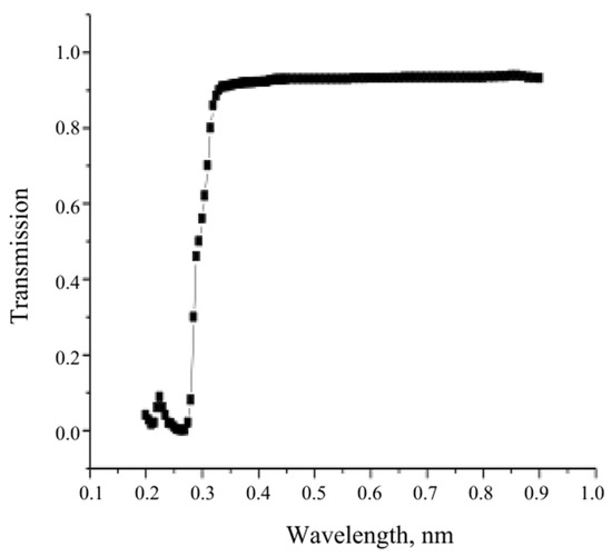
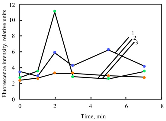
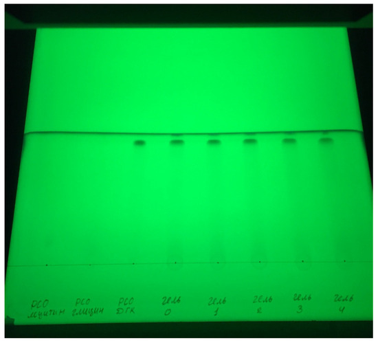
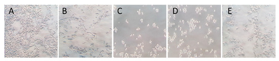
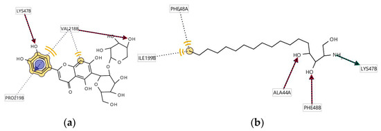
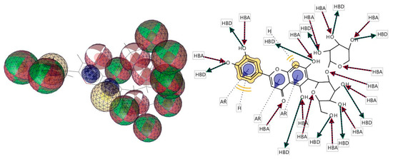
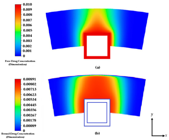
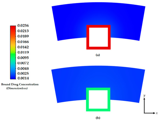
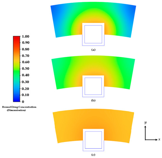
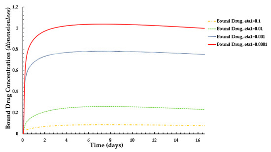
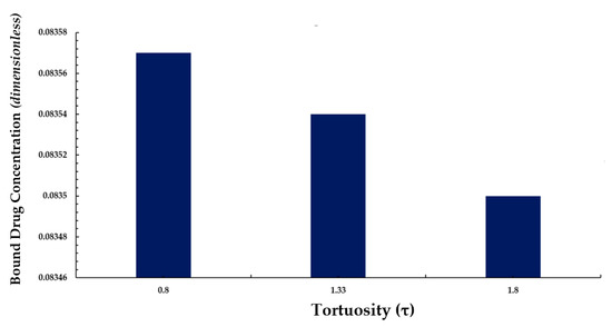
In modern dentistry, the problem of the prevention and treatment of peri-implantitis is relevant. Proposed methods of treating patients with peri-implantitis do not stop the pathological process with the possibility of achieving long-term remission. Liposomal complexes with dihydroquercetin make it possible to influence the pathogenetic links of the inflammatory process in periodontal tissues with the prospect of normalizing blood circulation and regeneration processes in the affected area. It has been established that the complex simultaneous effect of low-intensity laser radiation and a pharmaceutical (laserophoresis) provides the possibility of more significant penetration of the drug components into periodontal tissues. The study of the laserophoresis of the liposomal complex with dihydroquercetin in the treatment of patients with peri-implantitis is relevant. However, in the modern literature, there is a lack of studies on the effect of low-intensity laser radiation on the pharmaceutical structure of drugs based on the above-mentioned basis.
Full article
(This article belongs to the Special Issue Biomedical Optics)
►
Show Figures
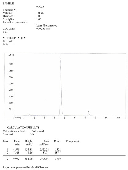
Figure 1

_copy_-_cópia.jpg)
.jpg)


