


Dent. J. 2023, 11(9), 208; https://doi.org/10.3390/dj11090208 - 31 Aug 2023
Abstract
►
Show Figures
The accuracy for the implant position transfer of a mounting fixture and a standardized open-tray implant level impression was compared. Ten aluminum master models with four implant analogs placed in different angulations were fabricated. By performing an open-tray implant level impression stone casts
[...] Read more.
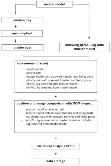
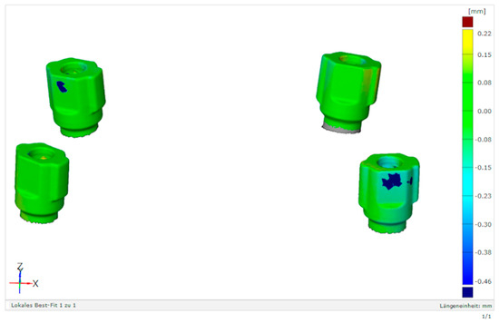
The accuracy for the implant position transfer of a mounting fixture and a standardized open-tray implant level impression was compared. Ten aluminum master models with four implant analogs placed in different angulations were fabricated. By performing an open-tray implant level impression stone casts were produced. The master models and stone casts were scanned (comparison group one) using a laboratory scanner. Deviations in the scan body surface were determined in the form of mean (absolute) point distances and (signed) surface distances. The same procedure was performed with a screwed transfer and by fixing the posts of the mounting fixture (comparison group two). The mounting device was applied to each master model and scanned in a fixed and detached state (comparison group three). In a point comparison, the open-tray implant level impression showed mean deviations of 43.6 µm and a mounting fixture of 44.6 µm with no significant differences (p < 0.05). There were significant differences between groups two and three. The angulation of the implants had no effect on the accuracy. In a surface comparison, the open-tray implant level impression showed mean deviations of 36.0 µm and a mounting fixture of 2.0 µm (p > 0.05). Within the limits of this study, the mounting fixture transferred the implant position with the same accuracy as the open-tray implant level impression with respect to point deviations.
Full article

Figure 1


.jpg)


