


by
, , , , , , , , , , and

J. Cardiovasc. Dev. Dis. 2023, 10(9), 375; https://doi.org/10.3390/jcdd10090375 (registering DOI) - 01 Sep 2023
Abstract
►
Show Figures
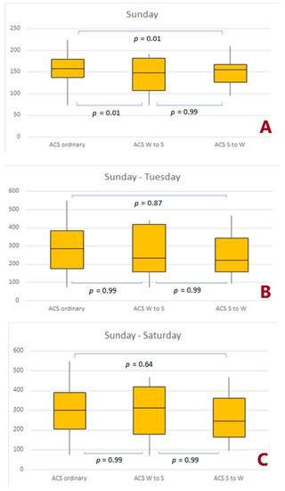
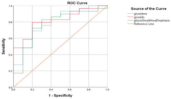
Introduction: Many factors related to the switch to summer/winter time interfere with biological rhythms. Objectives: This study aimed to analyze the impact of time change on clinical outcomes of patients with acute coronary syndromes (ACS) undergoing percutaneous coronary intervention (PCI). Patients and methods:
[...] Read more.
Introduction: Many factors related to the switch to summer/winter time interfere with biological rhythms. Objectives: This study aimed to analyze the impact of time change on clinical outcomes of patients with acute coronary syndromes (ACS) undergoing percutaneous coronary intervention (PCI). Patients and methods: Electronic data of 874,031 patients with ACS who underwent invasive procedures were collected from the Polish National Register of Interventional Cardiology Procedures (ORPKI) between 2014 and 2021. We determined the number of patients undergoing PCI and periprocedural mortality during the day of spring or autumn time change and within the first 3 and 7 days after the time change. Results: We demonstrated the impact of time changes on the periprocedural mortality of ACS patients within 1 day and the period of 3 and 7 days from the time change. We observed that the occurrence of all ACS and NSTEMI on the first day was lower for both time changes and higher in the case of UA and spring time change. The autumn time change significantly reduced the occurrence of all types of ACS. A significant decrease in the number of invasive procedures was found after autumn transition in the period from the first day to 7 days for ACS, NSTEMI, and UA. Conclusions: The occurrence of ACS and the number of invasive procedures were lower for both changes over time. Autumn time change is associated with increased periprocedural mortality in ACS and a less frequent occurrence of UA and NSTEMI within 7 days.
Full article

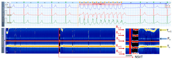
Figure 1


