


by
and

J. Dev. Biol. 2023, 11(3), 37; https://doi.org/10.3390/jdb11030037 - 31 Aug 2023
Abstract
Generating specialized cell types via cellular transcription factor (TF)-mediated reprogramming has gained high interest in regenerative medicine due to its therapeutic potential to repair tissues and organs damaged by diseases or trauma. Organ dysfunction or improper tissue functioning might be restored by producing
[...] Read more.
Generating specialized cell types via cellular transcription factor (TF)-mediated reprogramming has gained high interest in regenerative medicine due to its therapeutic potential to repair tissues and organs damaged by diseases or trauma. Organ dysfunction or improper tissue functioning might be restored by producing functional cells via direct reprogramming, also known as transdifferentiation. Regeneration by converting the identity of available cells in vivo to the desired cell fate could be a strategy for future cell replacement therapies. However, the generation of specific cell types via reprogramming is often restricted due to cell fate-safeguarding mechanisms that limit or even block the reprogramming of the starting cell type. Nevertheless, efficient reprogramming to generate homogeneous cell populations with the required cell type’s proper molecular and functional identity is critical. Incomplete reprogramming will lack therapeutic potential and can be detrimental as partially reprogrammed cells may acquire undesired properties and develop into tumors. Identifying and evaluating molecular barriers will improve reprogramming efficiency to reliably establish the target cell identity. In this review, we summarize how using the nematode C. elegans as an in vivo model organism identified molecular barriers of TF-mediated reprogramming. Notably, many identified molecular factors have a high degree of conservation and were subsequently shown to block TF-induced reprogramming of mammalian cells.
Full article
(This article belongs to the Special Issue Caenorhabditis elegans – a Model for Understanding Development and Disease)
►
Show Figures
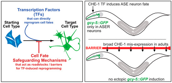
Figure 1


