


J. Imaging 2023, 9(9), 181; https://doi.org/10.3390/jimaging9090181 - 31 Aug 2023
Abstract
►
Show Figures
Rowing competitions require consistent rowing strokes among crew members to achieve optimal performance. However, existing motion analysis techniques often rely on wearable sensors, leading to challenges in sporter inconvenience. The aim of our work is to use a graph-matching network to analyze the
[...] Read more.
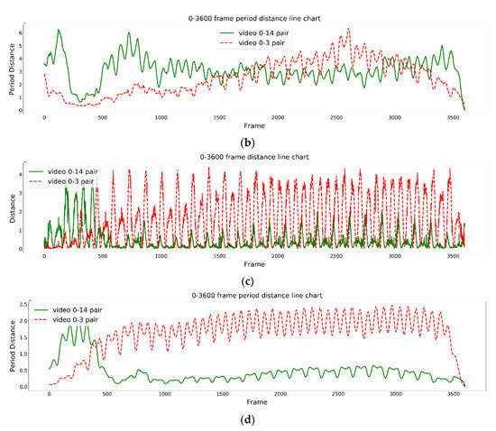
Rowing competitions require consistent rowing strokes among crew members to achieve optimal performance. However, existing motion analysis techniques often rely on wearable sensors, leading to challenges in sporter inconvenience. The aim of our work is to use a graph-matching network to analyze the similarity in rowers’ rowing posture and further pair rowers to improve the performance of their rowing team. This study proposed a novel video-based performance analysis system to analyze paired rowers using a graph-matching network. The proposed system first detected human joint points, as acquired from the OpenPose system, and then the graph embedding model and graph-matching network model were applied to analyze similarities in rowing postures between paired rowers. When analyzing the postures of the paired rowers, the proposed system detected the same starting point of their rowing postures to achieve more accurate pairing results. Finally, variations in the similarities were displayed using the proposed time-period similarity processing. The experimental results show that the proposed time-period similarity processing of the 2D graph-embedding model (GEM) had the best pairing results.
Full article
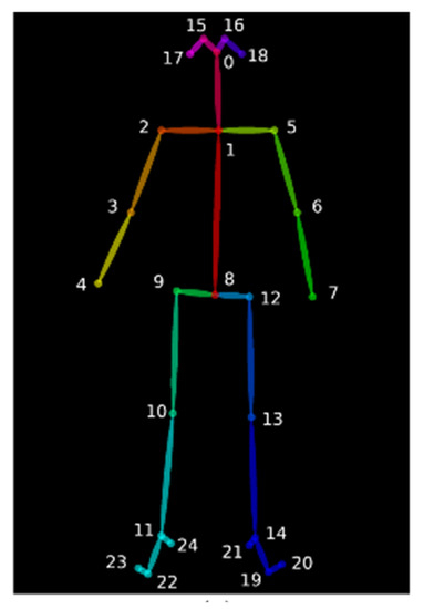
Figure 1


